Equipment


Digital Imagining Systems
With advances in digital imaging technology, we are now able to capture and document any part of the eye. At iSight, we have digital imaging systems for both the front surface (anterior segment) of the eye, and the inside (posterior segment) of the eye. Documentation of any feature of the eye allow us to closely monitor any changes or progression in eye diseases, or any abnormal features of the eye. The pictures will be saved to your file for comparisons in the future.
Anterior segment photography also allows us to fit specialty contact lenses (such as OrthoK lenses, or rigid gas permeable lenses) much more accurately, as we can see document how the lenses behave in the eye in between visits.
As the saying goes, "a picture tells a thousand words".
Optical Coherence Tomography (OCT)
Optical Coherence Tomography, or OCT, is an advanced technology used for retinal imaging and analysis. This technology provides highly detailed images of the retina as well as the optic nerve. These cross-sectional images are viewable in 2D and 3D, allowing for topographical mapping of the eye including retinal layer and nerve fiber layer thickness.
The accuracy of the OCT means that repeatability of measurement is very high and serial scans are very sensitive to changes in the eye that may signify the very earliest signs of eye disease such as glaucoma, macular degeneration, etc.
At Central Eye Care, we recommend a routine OCT scan if you have migh myopia (more than -6.00 D), or if you have any family history of eye diseases, such as glaucoma, or macular degeneration.
Remember, the earlier we detect any signs of abnormality, and the earlier we implement treatments, the better the clinical outcome of preventing vision loss.
Anterior segment photography also allows us to fit specialty contact lenses (such as OrthoK lenses, or rigid gas permeable lenses) much more accurately, as we can see document how the lenses behave in the eye in between visits.
As the saying goes, "a picture tells a thousand words".
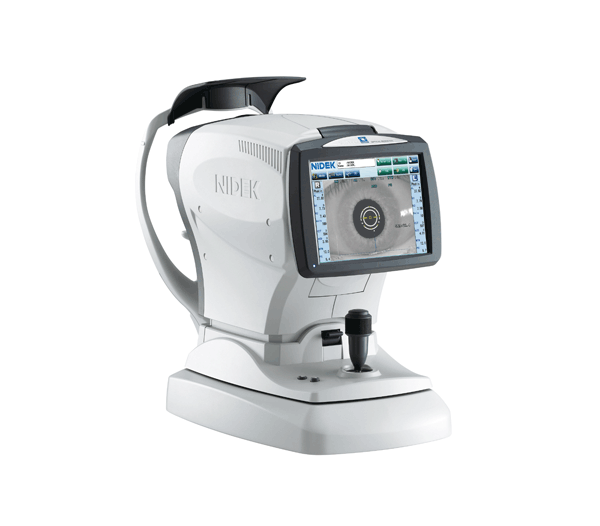
Corneal Topographer
A corneal topographer is a digital imaging technique for mapping the surface curvature of the cornea (the very front, clear part of the eye). The allows our Optometrist to evaluate the shape of the cornea before any contact lens fitting, and also to detect any corneal abnormality such as irregular corneal shape or eye diseases such as keratoconus.
At Central eye care, we routinely take images of the cornea before any contact lens fitting. This allows us to help you find the contact lens that best suits your eyes. (A common misconception is that contact lenses are a one-size-fit-all.) Not only will a properly fitted contact lens give you better comfort and vision, it will also protect the health of your cornea from poorly fitted lenses.
Corneal topography is also a MUST when fitting children with myopia control lenses, such as Orthokeratology fitting. This will ensure that each lens is custom made according to the natural curvature of the eye, and to monitor changes in the cornea during the treatment. Improperly fitted lenses without corneal topographer will lead to serious consequences.
Intelligent Refractor RT-6100
-
Breakthrough refractor for precise and efficient examinations
-
Streamlined refractor head
-
User-friendly control console
-
Binocular open refraction
-
Program edit function
-
Simplified data transfer
Optical Biometer AL-Scan
-
Effortless measurement of 6 clinical parameters in 10 seconds
-
Anterior segment observation with Scheimpflug imaging and double mire ring keratometry
-
Optional built-in ultrasound biometer
-
IOL power calculation and IOL constants optimization
-
Additional features with AL-Scan Viewer for NAVIS-EX
Tonometer
Tonometry is a test to measure the pressure inside your eyes. The test is used to screen for glaucoma.
How the Test is Performed
There are many methods of testing for glaucoma.
The most accurate method measures the force needed to flatten an area of the cornea.
-
The surface of the eye is numbed with eye drops. A fine strip of paper stained with orange dye is held to the side of the eye. The dye stains the front of the eye to help with the exam.
-
The slit-lamp is placed in front of you. You will rest your chin and forehead on a support that keeps your head steady. The lamp is moved forward until the tip of the tonometer just touches the cornea.
-
Blue light is used so that the orange dye will glow green. The health care provider looks through the eyepiece on the slit-lamp and adjusts a dial on the machine to give the pressure reading.
-
There is no discomfort with the test.


The OCULUS Keratograph 5M
The OCULUS Keratograph® 5M is an advanced corneal topographer with a built-in real keratometer and a color camera optimized for external imaging. Unique features include examining the meibomian glands, non-invasive tear film break-up time and the tear meniscus height measurement and evaluating the lipid layer.

